Customer Case Study #3
Reducing the burden on technicians
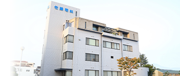
Medical corporation Seiyaku-kai Sato Eye Clinic

Sato Eye Clinic in Kojima, Kurashiki, Okayama Prefecture, has been a dedicated eye care facility since its founding in 1886 (Meiji 19). For over 130 years, the clinic’s successive directors have honed their skills and knowledge at major hospitals throughout Japan, continuing a proud tradition of providing the latest in ophthalmic care. In addition to improving the standard of medical care in the region, the clinic also strives to train the next generation of healthcare professionals and establish systems to safeguard children’s eye health.
Dr. Shinya Watanabe, the clinic’s Vice Director, shared insights on the impact of introducing GAP and the changes it has brought to the clinic’s operations.

■ Key Points of This Case Study
- ― Operated alongside the Humphrey Field Analyzer
- ― Immediate testing available without an appointment, reducing the burden on patients
- ― Fewer testing steps and time savings allow technicians to handle other tasks
Key Factors for Product Introduction
At our outpatient clinic, the proportion of patients with glaucoma is particularly high. This has led to challenges such as longer waiting times for visual field tests and difficulties in securing appointment slots due to congestion in the visual field testing schedule. Originally, we had only one Humphrey Field Analyzer, but after learning about GAP at an academic conference, we decided to introduce it as a visual field testing device with shorter testing times, to be used in parallel with the Humphrey Field Analyzer. For patients who have not yet been definitively diagnosed with glaucoma, the Humphrey Field Analyzer can take a considerable amount of time. Therefore, we were looking for a device that could provide a quick examination.
Another key factor was that GAP easily exports images to our electronic medical records and is compatible with the visual field analysis software we use for longitudinal assessments. This level of integration with our electronic medical records was also a crucial point for us.
Challenges and adjustments made in the testing operations when introducing GAP
For a while, we didn’t change the number of appointment slots for visual field tests. Instead, we switched to using GAP testing for patients with mild cases to observe the testing time in actual clinical practice, minimizing confusion and burden for the technicians.
At our clinic, we perform OCT on all patients over 40 years old, as well as those with high myopia or optic disc cupping. For patients with abnormalities found on OCT or other tests who hadn’t yet undergone visual field testing, the availability of GAP allowed us to conduct immediate visual field tests without an appointment. This helped reduce the number of patient visits and lessen their burden.
Initially, we expected to mainly use the eye-tracking feature, which is a selling point of GAP. However, in cases where patients have difficulty keeping their eyes open, the examination time can actually become longer. Depending on the patient’s condition, we switch between the conventional fixation mode (which uses button operations) and the eye-tracking mode to ensure the examination is completed efficiently.

Benefits and improvements observed since introducing GAP
Unlike conventional visual field testing devices, GAP has several advantages:
- There is no need to replace corrective lenses when switching eyes; aligning the patient’s eye position is all that’s required, making the process much simpler.
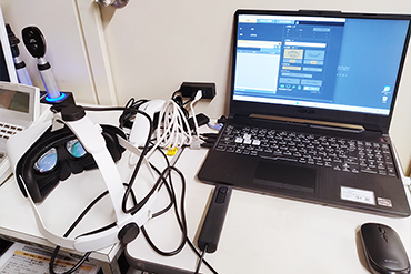
- In the past, we needed to support the patient’s body to ensure their head didn’t move away from the device. However, with the new goggle-type design, there’s no need to monitor head movement, which has reduced the burden on technicians.
- Preoperative testing for cataract surgery can now be completed in about one minute with GAP’s screening test.
These factors have all contributed to reducing the burden on technicians. I believe these benefits are particularly significant for eye clinics that specialize in glaucoma care.
Patient Reactions to GAP

In ophthalmology, there are many testing devices with various designs, and GAP’s use of a head-mounted display gives it the appearance of the latest medical technology, which patients perceive very positively.
Moreover, the eye-tracking feature can capture the attention of children who might struggle to concentrate, making it easier to complete the test by engaging them in a way that feels like playing a game.
Patients also seem to feel that the overall testing posture and burden are lighter compared to conventional visual field testing devices. As a result, we have the impression that fewer patients find the examination physically or mentally challenging.
Expectations for Future Developments of the GAP
At our clinic, we use conventional visual field testing devices for patients with clearly defined visual field defects from glaucoma. For patients over 40 years old or those requiring glaucoma screening, such as those with optic disc cupping, we use GAP. As such, in GAP testing, accurately distinguishing whether visual field defects are present in mild or borderline cases is particularly important.
We look forward to the development of diagnostic support software that can automatically determine whether abnormalities are present by referencing glaucoma progression patterns and filtering out obvious testing noise from the results.
Dr. Watanabe, Thank you for your cooperation in the interview.
Medical corporation Seiyaku-kai Sato Eye Clinic
Address:
1-88, Kojima Ekimae, Kurashiki city, Okayama prefecture